

Refer to the drawing of bone on demonstration and identify Haversian canal systems, (osteons), which are concentric rings of bone surrounding a canal. The forming of this complex structure is called ossification. Name some replacement bones seen in this week’s lab.Įxamine the microscope demonstration of dry ground bone. The axial skeleton is derived from sclerotome (epimere), the appendicular skeleton from somatic hypomere. Cartilage must be removed before bone can be deposited. To widen it, the periosteal membrane directly secretes perichondrial bone in strips. New cartilage made by chondroblasts grows mainly at the ends and at epiphyses to lengthen the bone. Replacement bones ( endochondral bones) are formed by replacing embryonic hyaline cartilage with bone. Name some dermal bones seen in this week’s lab. Other membrane bones are deep bones that also do not have a primary cartilage matrix. Dermal bones are plate like, but can become thicker or grow at the edges. Osteoblasts form a calcium phosphate matrix and deposit salts, then become osteocytes.
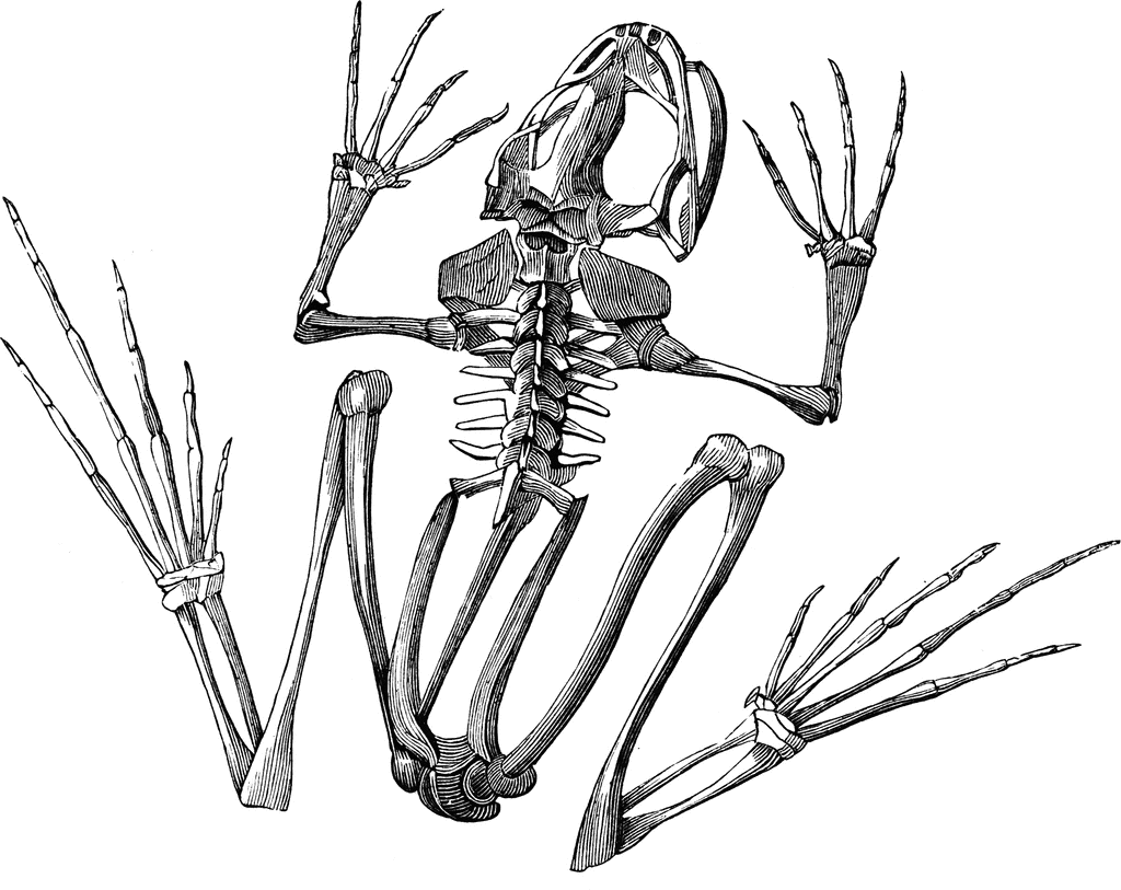
#SKELETON SYSTEM OF FROG SKIN#
ĭermal bone forms directly in the dermis of the skin from mesenchyme. Fibrous tissue from the newly discovered syndetome layer (between myotome and sclerotome) makes up tendons, which join bone and muscle together and ligaments, which join bones to each other.īone can either be intramembranous (membrane bone) (eg: dermal bone) or replacement. General connective tissue, from embryonic mesenchyme is dispersed throughout the body and also is found as adipose (fat) and areolar tissue and as blood plasma and lymph. Name and draw the three major types of cartilage. Cartilage is derived from mesenchyme and forms a matrix for bones, ends of bones, intercostals from ribs, nose, ears etc. They have different amounts of calcification, which is the addition of calcium salts, and differ in their fibre content. Note the different types of cartilage on display. All nourishment is by means of diffusion through the matrix, so cartilage seldom acquires much thickness although it grows by internal expansion. No blood vessels or nerves penetrate the matrix. These cells (chondrocytes) on the underside of the perichondrial membrane secrete the matrix. Notice the nuclei of discrete cells in lacunae (spaces) embedded in a matrix of polysaccharides. 1) Be able to identify the 3 major types of cartilage and explain where they are prevalent in the body.Ģ) Be able to differentiate between the 2 major types of bone, describe how they are formed, and where they are located in the body.ģ) Identify the additions to vertebrae and their function.Ĥ) Identify the different types of vertebral centra and the vertebrate groups they occur in.ĥ) Understand regional specialization of vertebrae in the different vertebrate groups.Ħ) Explain the form and function of the pelvic and pectoral girdles and determine which bones are dermal and which are replacement bones.ħ) Identify the major bones in the limbs.Ĩ) Give examples of how limbs are adapted for different functions.Įxamine the microscope slides of hyaline cartilage on demonstration.


 0 kommentar(er)
